shared facility for electron microscopy an x-ray microanalysis (SCMEM)
Technical Manager : Andrei LECOMTE
Scientific Manager : Jean CAUZID

Summary

The SCMEM – Shared Facility for Electron Microscopy and X-Ray Microanalysis
The SCMEM – Shared Facility for Electron Microscopy and X-Ray Microanalysis is:
♦ part of the RéGEF network,
♦ and in the process of being labeled Infra + via the ANATELo platform.
GeoRessources is responsible for managing the SCMEM, the Université de Lorraine’s long-standing Shared Electron Microscopy and X-ray Microanalysis Service. Located in Nancy, the SCMEM is open to members of Université de Lorraine and to external parties, including academic and industrial institutions.
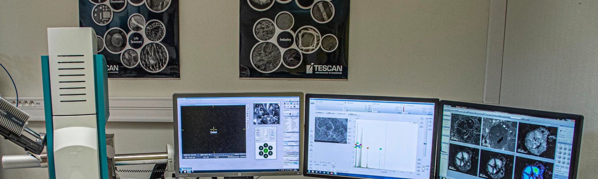
The SCMEM offers a range of equipment based on electron-matter and X-ray-matter interactions. They allow the observation and characterization of solid samples at very high magnifications.
It is possible to obtain:
♦ 2Topographical information about the sample (secondary electron imaging, SE),
♦ Chemical contrast (back-scattered electron imaging, BSE),
♦ Cathodoluminescence images,
♦ Elemental distribution images (X-ray mapping),
♦ Quantitative X-ray analyses at the micrometer scale,
♦ Particle analyses (size, shape, composition, etc.),
♦ Mineralogical characterization (mineral mapping, phase distribution, MLA – Mineral Liberation Analysis)

Technical expertises


What are the technical areas of expertise of the Shared Electron Microscopy and X-ray Microanalysis Service (SCMEM) platform?
♦ Characterization of materials using scanning electron microscopy (SEM)
♦ Elemental mapping and X-ray microanalysis (SEM, microprobe, µXRF)
♦ Automated mineralogy using SEM/µXRF
♦ High-resolution imaging
See all the expertise of Georessources in Microscopy and spectroscopies.
Equipments and fundings
The SCMEM has a wide range of imaging and X-ray analysis tools:
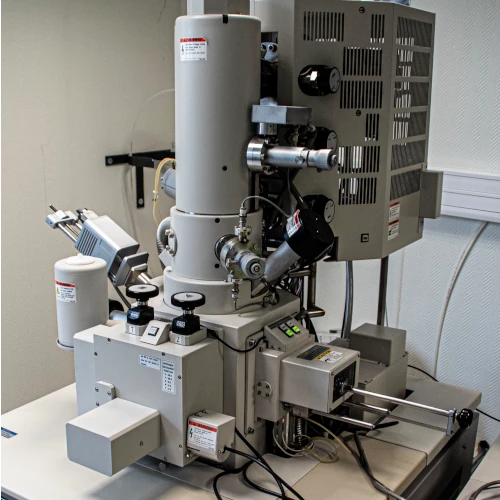
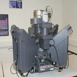
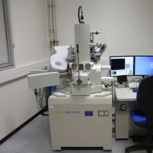
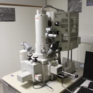
♦ A HITACHI S4800 field emission scanning electron microscope (SEM) (cold cathode, Cold-FEG) dedicated to very high magnification imaging. It is coupled with an energy-dispersive X-ray spectrometer (EDS-SDD) and can be used in transmission mode (bright field/dark field) thanks to a STEM detector.
♦ A JEOL JSM-7600F field emission SEM (Schottky-FEG) dedicated to quantitative analysis thanks to a 20mm2/WDS Oxford Instruments EDS coupling.
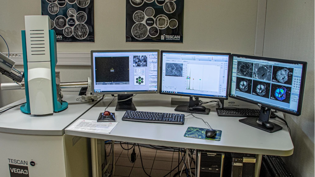
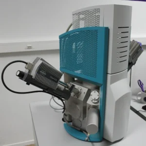
♦ A conventional SEM (tungsten filament) TESCAN VEGA 3 LM, coupled with two Bruker XFlash6 30mm2 EDS detectors and a Gatan ChromaCL2UV cathodoluminescence system. It is possible to work at partial pressures up to 150 Pa (500 Pa with diaphragm change).
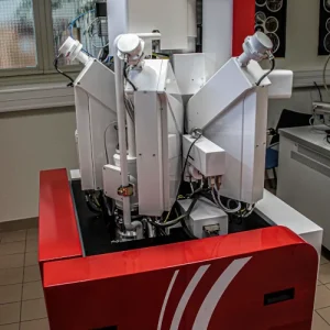
♦ A CAMECA SXFive TACTIS Castaing microprobe (electron probe microanalyzer, EPMA) equipped with a LaB6 tip, five vertical spectrometers, including an extended spectrometer, and a cathodoluminescence device.
♦ A CAMECA SX100 Castaing microprobe equipped with a W gun and five vertical WDS spectrometers.
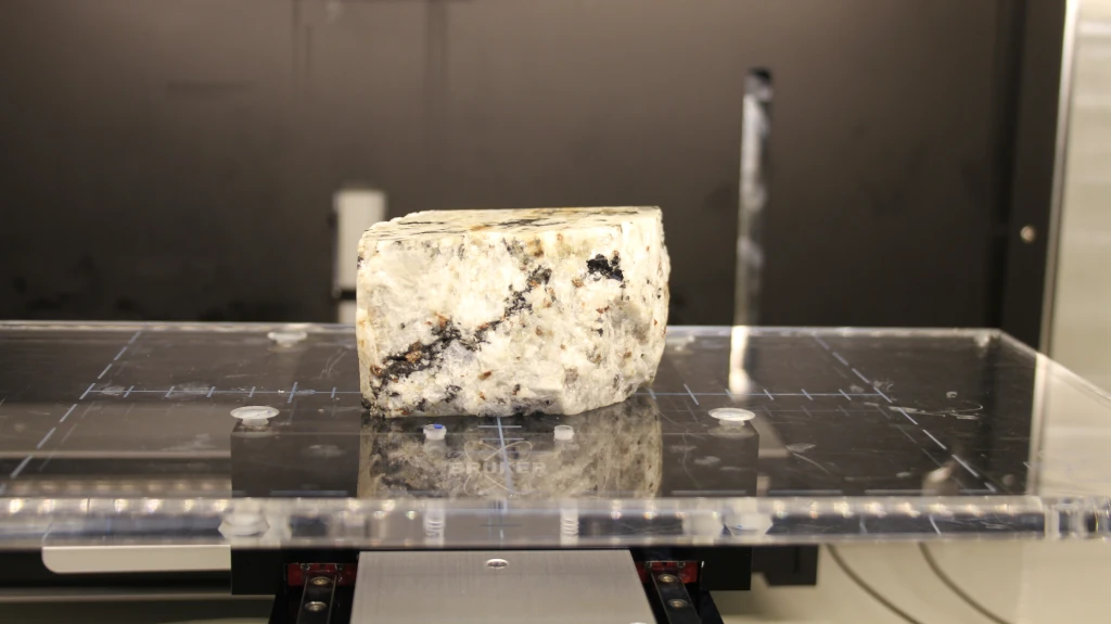
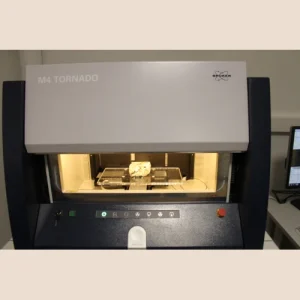
♦ A BRUKER M4 TORNADO µ-XRF equipped with a Rh tube and two 30 mm² EDS detectors.
The SCMEM also offers access to automated mineralogy thanks to the Bruker AMICS solution available on SEM or µXRF, depending on the objectives of the study and the type of samples.
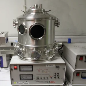
Finally, the department has two vacuum evaporators, JEOL JEE-420 and CRESSINGTON 308R, used for sample metallization.
How to get to the Shared Facility for Electron Microscopy an X-ray Microanalysis
The Shared Facility for Electron Microscopy an X-ray microanalysis (SCMEM) is located at the following address:
Faculté des Sciences et Technologies de Nancy
SCMEM
Gate 5c
54500 Vandoeuvre-lès-Nancy
GPS coordinates: 48.6642191121907, 6.159416501041887
Contact
If you would like to contact GeoRessources, please feel free to send us a message by clicking on the link below and filling out the form.
