X-Ray Tomography
Contacts:
► Christophe Morlot, technical manager
Tel. +33 (0)3 72 74 55 01
Mail : christophe.morlot@univ-lorraine.fr
► Jérôme Sterpenich, scientific manager
Tel. +33 (0)3 72 74 56 59
Mail : jerome.sterpenich@univ-lorraine.fr
The platform in video
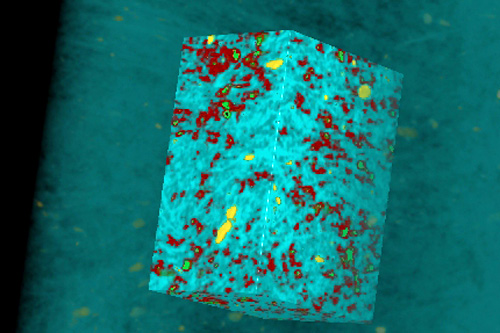
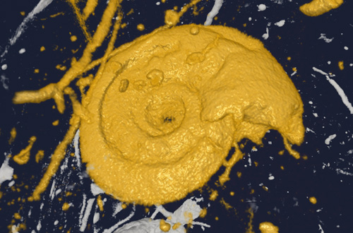
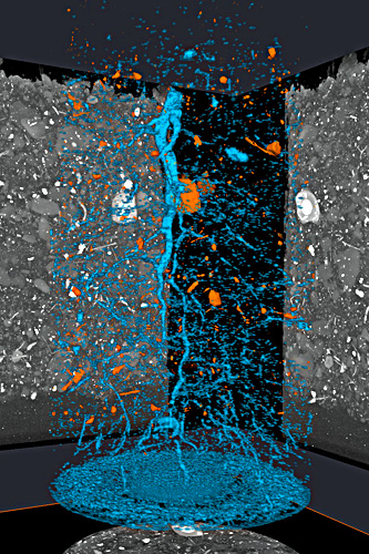
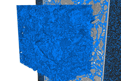
The GeoRessources Nanotom Phoenix X-ray tomograph (GE) is used to explore the architecture of solid samples at resolutions of less than micrometer-scale. Samples are illuminated with an X-ray beam which allows us, using an X photon detector, to record the adsorbed beam that has passed through the solid to the analyser. Image acquisition is repeated at different rotation angles and the different sections are then reconstructed using algorithms to form a three-dimensional image of the sample.
This non-destructive technique provides us with the capability to examine the internal structure of a sample: dimensions, shape, the spatial distribution of elements in relation to one another, and sample heterogeneities and defects (e.g., pores, inclusions and mineral phases). The high-quality 3D imagery allows the reconstructed specimen to be manipulated in all spatial directions and virtual sections can be obtained through any part of a sample.
Specimen sizes range from less than 1 millimetre up to a maximum of few tens of centimetres. A spatial resolution of 0.5 μm can be achieved for the smallest samples.
|
Characteristics of the X-ray tube
|
 |
|
Computer resources
Dell and HP workstations Dedicated cluster models 3D-modelling software: VGStudio 2.2 and Avizo 9.4 |
 |
fees 2019
per sample
Academic partner user: 261 € HT
Academic user not a partner: 385 € HT
Private user: 432 € HT
Academic users: these are users of EPSCP, EPST and related research units. This rate is also applicable to CREGU.
|
|
  |
