Optical microscopy
Contacts:
► Karine Pistre, technical manager
Tel. +33(0)3 72 74 55 02
► Alexandre Tarantola, scientific manager
Tel. +33(0)3 72 74 55 67
► Cédric Carpentier, scientific manager
Tél. +33 (0)3 72 74 55 51
The platform in video
Dedicated to the characterisation of microscopic objects, the platform comprises several types of equipment:
- Transmitted and reflected light optical microscopes coupled to high-resolution cameras for the study of thin sections and polished sections.
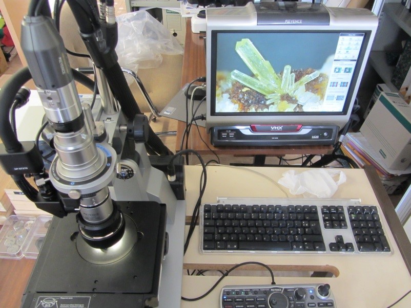
- A digital microscope, for sharp observation of objects in two and three dimensions, thanks to a depth of field 20 times greater than that of a conventional optical microscope.
- Microscopes equipped with cathodoluminescence or ultraviolet fluorescence systems for the identification of defects, cracks, impurities, organic inclusions and growth zones, allowing the establishment of paragenetic sequences.
- Microscopes equipped with microthermometry stages for measuring the temperatures of phase changes in fluid inclusions, permitting the salinity of fluids and their minimum trapping temperatures to be determined.
- A microscope equipped with a system for measuring the reflectance and fluorescence of organic matter in order to assess the degree of thermal maturation.
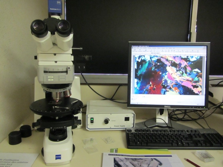
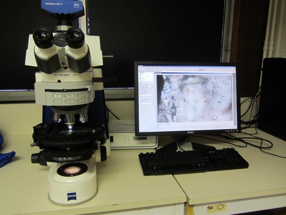
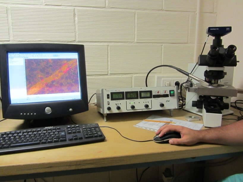
Optical microscopy - transmitted and reflected lightThe GeoRessources Laboratory provides three optical microscopes for the observation and analysis of different geological specimens including thin sections, thick sections and polished sections. Description and applications: |
 |
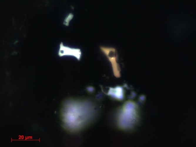
Zeiss AxioImager A1m optical microscope equipped with a UV lampThe principle of this technique is based on emission of visible light by a specimen that has been excited using a higher-energy light (with a lower wavelength) such as UV light. Description of the microscope: - Sample illumination with reflected UV light Applications: |
Inclusions fluides hydrocarbonées, Yemen
Inclusions fluides hydrocarbonées, Mexique |
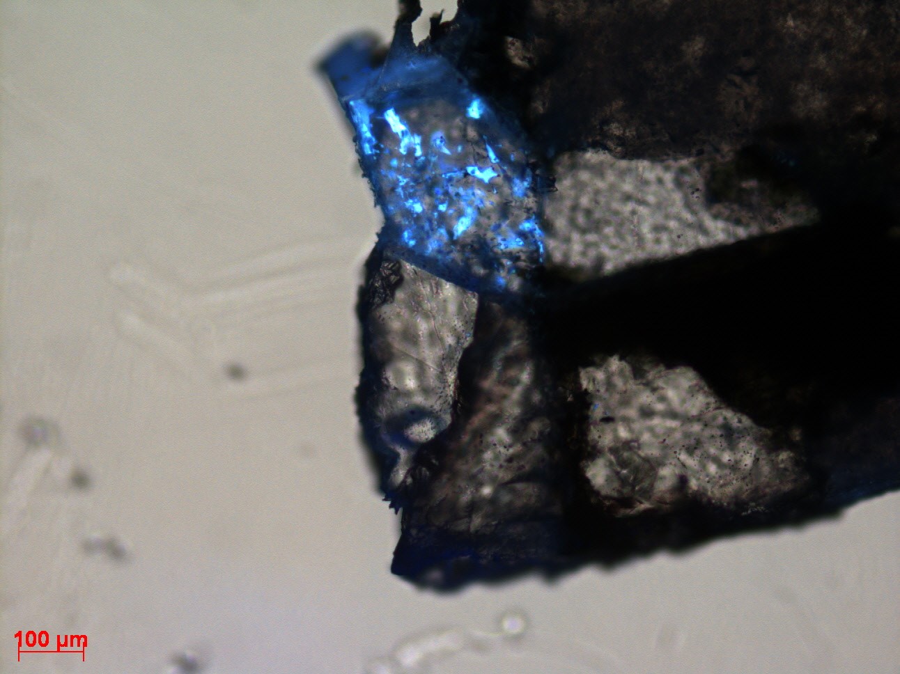
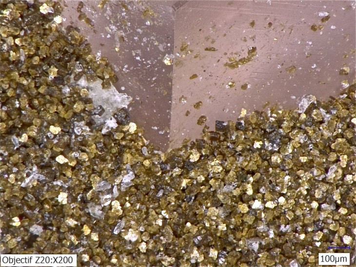
Keyence VHX – 1000 digital optical microscopeThe Keyence VHX-1000 digital microscope is a multi-use instrument that provides a depth of field that is 20 times greater than that of a conventional optical microscope, enabling sharp observations of 3D objects. Its comprehensive set of objectives (20x-200x, 100x-1000x, 500x-5000x) and adjustable observation system (illumination by inclined reflected light or transmitted light) allow us to obtain high-quality images of a large variety of samples, including thin sections, polished sections and macroscopic samples. Applications: |
Chlorite sur fluorine, Alpes
Plans nets d’inclusions fluides, Alpes |
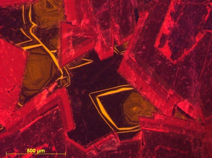
Microscope equipped with a cold-cathodoluminescence systemCathodoluminescence is defined as the emission of light photons by a solid surface that has been bombarded with electrons (emitted from a cold cathode). The technique is non-destructive; however the impact of the electron beam can cause alteration of the minerals or paste. This can lead to the creation or ionization of defects, or may trigger their diffusion. The wavelength of the photons depends on the nature of the minerals and is generally in the visible portion of the spectrum although can also be in the infrared (IR) or ultraviolet (UV) portions. Applications: |
Dolomite et calcite, Pyrénées |
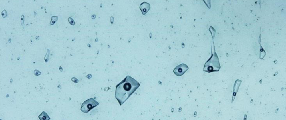
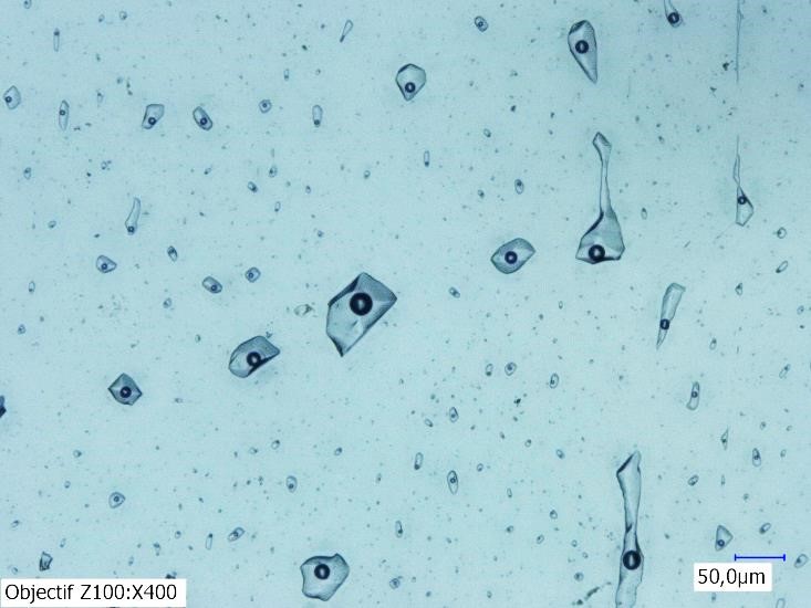
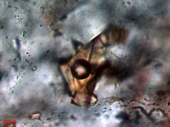
Microthermometry of fluid inclusions
Fluid inclusions are small, fluid-filled cavities inside a mineral. Fluids are trapped during growth of the crystal or after crystal formation. Fluid inclusions may contain several phases that can be observed under the microscope: a liquid phase (e.g., H2O, petroleum, or a CO2 or H2S dense phase), gas phase (e.g., water vapour, CO2, CH4, N2, H2S, H2 or O2) or solid phase (e.g., chlorides, carbonates, silicates, sulphates). Microthermometry is an analytical technique used to identify and characterise the fluids that have circulated in rocks by measuring the temperatures at which phase changes occur in the interior of the fluid inclusions. - Cooling (to -180°C): nucleation phase of ice and salt or gas hydrates. Description of the systems: Applications: - Determining the nature of dissolved salts by measuring the eutectic. |
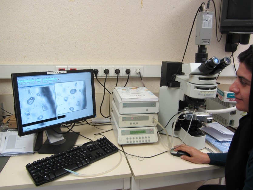 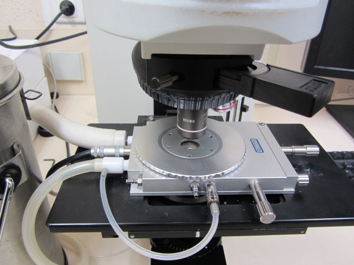
Platine Linkam THMS600
Inclusion fluide hydrocarbonée, Yemen |
Confocal microscopyNikon TE2000-U inverted laser scanning confocal microscopeConfocal microscopy is a high-resolution imaging technique based on laser excitation of a self-fluorescent sample. The excitation produces the emission of fluorescent rays from the different planes of the specimen. A diaphragm is placed in front of a detector (confocal aperture) enabling fluorescence emitted by planes located outside the focal plane to be eliminated. The fluorescent rays then pass through filters that separate the excitation and emission wavelengths, before arriving at the detection system. Electron photomultipliers record the photon emission and transform the light signal into a 2D image that corresponds to the specimen section. A three-dimensional image is obtained by simultaneous displacement of the stage along the Z-axis. With minimal alteration and preparation of the sample, we are therefore able to obtain extremely fine optical sections (from 0.5µm thickness) using a pinhole, and to achieve a true optical sample dissection. Description of the system: Advantages and applications: |
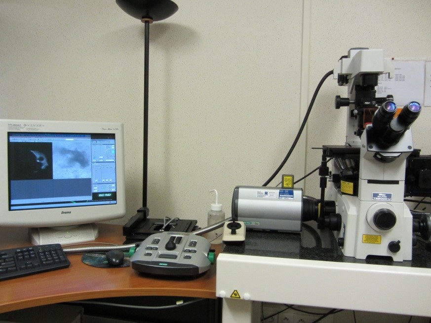 |
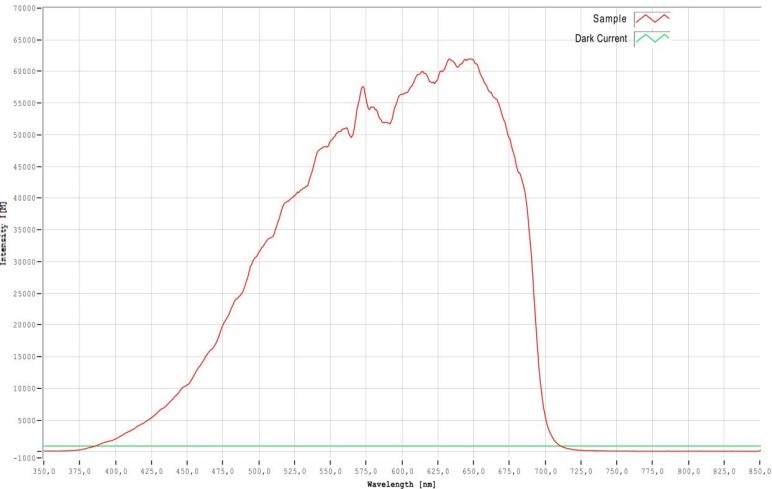
Optical microscopy of organic matterZeiss AxioImager microscope with system for measuring fluorescence and vitrinite reflectanceCoals are classified according to their rank, which reflects their enrichment in carbon during the burial process. The variation is mainly determined by an increase in temperature, from peat, which marks the first stage of thermal maturation (diagenesis), up to graphite, which marks the first stages of metamorphism. Macerals (vegetal matter), the main constituents of coal, are divided into three groups: vitrinite, leptinite and inertinite. Vitrinite is the most abundant of these. Description of the system: Applications: |
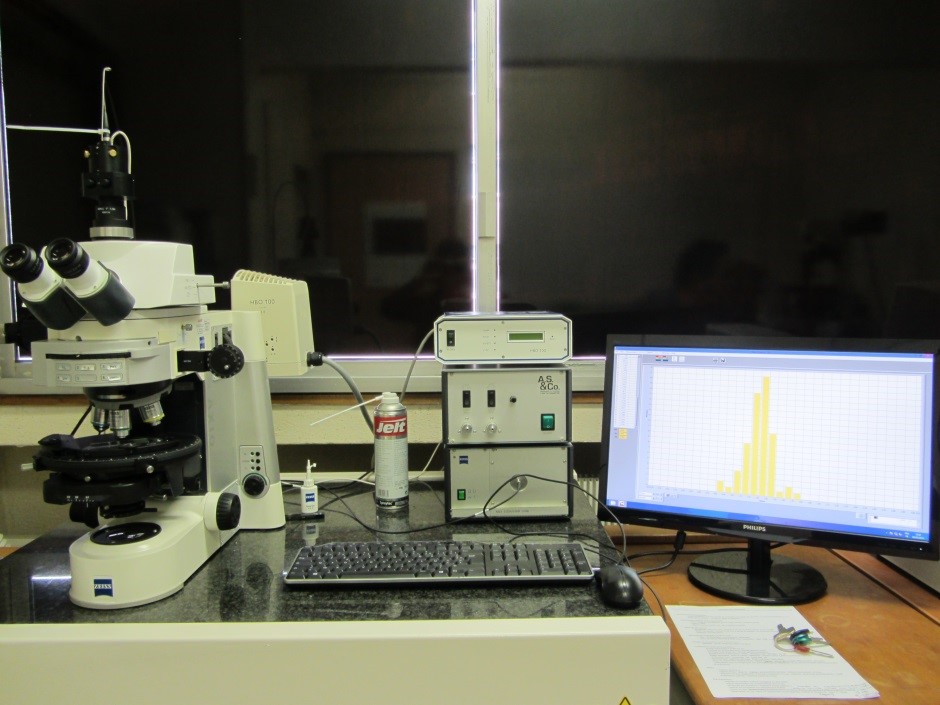 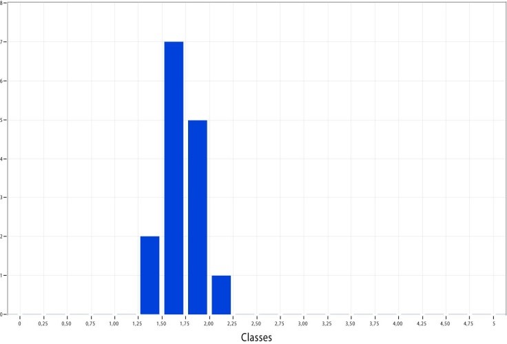
Histogramme de réflectance d’un alum shale, Suède Spectre de réflectance d’un alum shale, Suède |